|  Anthropomorphic
Molded Around Skull
Checks Important Physical Parameters
Provides Realistic System Check
Ideal For Training
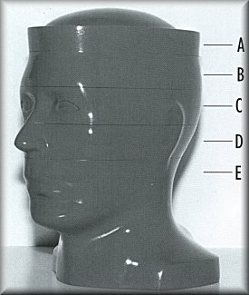
Polycarbonate Base Dimensioned For Scans Parallel or Perpendicular to Longitudinal Axis
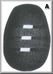
SECTION A: Aluminum plates, 0.4mm thick, set at a 45° angle, measure beam width and slice thickness.
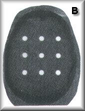
SECTION B: The dosimetry section provides patient-exposure controls, and checks on abnormal internal doses before
actual patient scans. Customization to accommodate TLD with rods or chips is available.
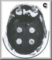
SECTION C: Cylindrical tumors of graded sizes and radiodensities establish high and low-contrast resolution and
demonstrate partial beam-averaging effects.
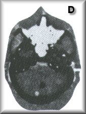
SECTION D: The anthropomorphic section provides actual auditory ossicles in an air-contrast inner ear and a
fractured petrous bone. Small tumors in the posterior fossa are placed in an area which most scanners image with
difficulty.
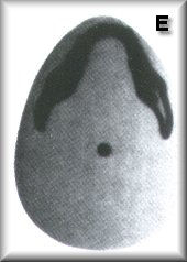
SECTION E: 1/4 Inch diameter aluminum rod on the phantom's longitudinal axis, checks general alignment, and
"DryWater" gives a realistic "noise check."
|


