 |
|
|
|

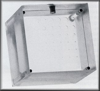
| Digital Stereotactic Breast Biopsy Accreditation Phantom. |
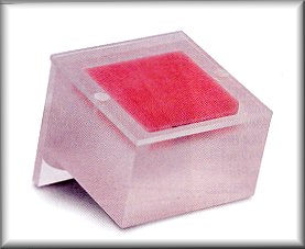 | 
The Problem In the past, there was not an easy way to compare the image quality of conventional and digital biopsy mammography units, because the field of view on the digital system is typically much smaller than the 24 x 30 cm field of view on conventional mammography units. In order to image the Mammographic Accreditation Phantom (specified by the ACR) on the biopsy units, the user has to move the phantom to various positions in order to obtain four separate images, to be sure all objects were imaged. This is a very inconvenient, time consuming task. Problem Solved! How? The small size of the phantom permits fast, easy comparison of conventional and digital image quality, because you can attain an image of the entire unit in a single exposure! The objects are some of the same ones found in the Mammographic Accreditation Phantom specified by the ACR, so it makes comparison of the two imaging systems easy. Weight: 1.2 kg (8.7 oz.) *Designed by Carol Mount, R.T. (R) (M), and Joel E. Gray, Ph.D., Department of Diagnostic Radiology, Mayo Clinic, Rochester, MN 55905. Manufactured under licensing agreement with Mayo Foundation for Medical Education and Research. |
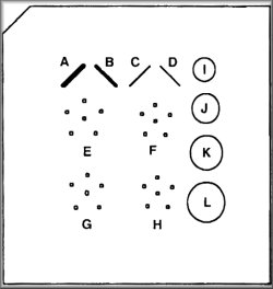 |
| Contrast Detail Phantom For Mammography. |
  |
The 18-227 Phantom for Mammography is designed to provide a means of quantitatively testing and monitoring the total performance of an entire mammographic imaging chain. Its small size, as well as the number and distribution of holes simulating embedded objects, make this phantom particularly useful in evaluating digital spot mammography systems. With 49 holes generating subtle contrast variatons, the phantom makes it possible to detect small changes in overall system performance. This Contrast Detail Phantom contains a 7 x 7 matrix of objects. The diameter of each row of objects decreases from 0.169" to 0.007". In each row, the subject contrast decreases from approximately 6.6% to 0.41% at mammographic energies. This Phantom is easy to use....Simiply place the phantom on the image receptor surface in the same positon as a breast. Position the x-ray tube and compression device as in a craniocaudal examination. When using the phantom on prone-position breast biopsy systems, use the rotating top plate of the phantom against the image receptor. Choose the appropriate kV and mAs factors (26kV and 60 mAs works well on most systems), or select automatic exposure control. |
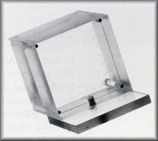
| Contrast Detail Mammogrraphy (CDMAM) Phantom. |
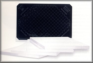 |
CDMAM PHANTOM
The 18-227 CDMAM (Contrast Detail Mammography) Phantom was developed to evaluate conventional mammographic x-ray equipment, film, and cassettes. However, with the rise of digital imaging in mammography, expecially when performing stereotactic breast needle biopsies and pre-operative needle localizations, the phantom can aid in achieving improved image quality, processing, display, and speed in these new modalities! Quality control of the technical aspects of mammographic equipment usually is performed by measurement of the physical parameters of the x-ray equipment, screen-film combination, developing process, and observation conditions. However, the main issue in quality control should be the assurance that absolutely correct information (with as much |
| anatomic detail as possible) about the tissue under examination, be transferred to the radiologist! To
facilitate this, the CDMAM was developed! What Makes the CDMAM Phantom So Special? Nuclear Associates' CDMAM Phantom consists of an aluminum base with gold discs (99.99% pure gold) of varying thicknesses and diameters, which is attached to a plexiglass cover. The 5 mm thick plexiglass cover (PMMA plate) has a 2 mm deep cavity which accomodates the aluminum base with gold discs. The assembly (PMMA + aluminum) has a plexiglass equivalent thickness of 10 mm, under standard mammography-exposure conditions. The aluminum base is .05 thick Al 1050 (99.5% pure aluminum). The base has been polished and anodized black. Precisely measured gold discs of varying thickness (range = 0.05 to 1.60 um) and diameter (range = 0.10 to 3.20 um) have been attached to the base by means of evaporization. Finally, the base has been airbrushed to protect the gold discs. The "Gold Standard" of Mammographic Phantoms! The discs are arranged in 16 rows and 16 columns. Within a row, the disc diameter is constant, with logarithmical increasing diameter. The precision of the disc diameter and thickness makes the CDMAM Phantom an ideal tool for conducting contrast-detail and other image quality experiments. A line pattern has been engraved onto the plexiglass cover and treated with aluminum containing paint. The x-ray image will show a number of squares ordered in 16 columns and 16 rows, with the disc diameter shown for each row, and the disc thickness for each column. About the "Gold Standard" CDMAM Phantom Nuclear Associates' CDMAM Phantom includes a set of 4 plexiglass plates, which are used for the simulation of different |
|
| breast thicknesses. The plates are 10 mm thick and the same dimensions as the phantom. The plates are marked in one
corner, for identification of the configuation of plexiglass and phantom in an x-ray image. The phantom and plexiglass
plates match the standard mammography film size (18 x 24 cm). Under standard mammography-exposure conditions (Mo-anode, 30 um Mo-filtration, 28 kV), the phantom has a plexiglass equivalent thickness of 10 mm. The actual attenuation of the CDMAM Phantom depends on the configuration of the phantom and plexiglass plates. The effective energy of the phantom-plane will be higher when more plexiglass is added to the top and bottom of the phantom. Using the CDMAM Phantom is Easy! To make an x-ray image, the CDMAM Phantom should be positioned on the bucky with the smallest disc diameters at the thorax side, in combination with one or more plexiglass plates. The markings on the plexiglass plates should be aligned at the thorax side of the bucky. On digital sterotactic systems with smaller fields of view, specific portions of the phantom can easily be imaged as well. The density of the image has to be checked after the film has been processed. In a series of CD images, all images should approximately have the same densities in a reference-position on the film. *The CDMAM Phantom is a result of extensive research by M.A.O. Thijssen, PhD., K.R.Bijkerk, MSc, and J.M Lindeyer, BSc., Project: Quality Assurance in Mammography (QAMAM), Department of Diagnostic Radiology, University Hospital Nijmegen, St. Radboud, The Netherlands. |
 |
|