Pacific Northwest X-Ray Inc.

       Nuclear Medicine Heart/Thorax Phantoms.       


Radiology Support Devices! 
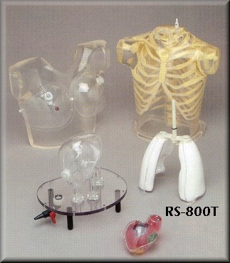
HEART/THORAX PHANTOM (Includes items RS-801 through RS-810)
The Heart/Thorax Phantom is ideal for evaluation of detectability, extent and severity of myocardial infarets in patients. This Phantom also provides valid assessment of mammoscintigraphy techniques. The Striatal Phantom optimizes quantitative imaging in patients, using PET or SPECT.

  • Heart/Thorax Phantom for Cardiac SPECT/PET and Mammoscintigraphy
  • Myocardial perfusion SPECT is a widely-used, non-invasive method for the diagnosis and management of patients with coronary disease. However, nonuniform photon attenuation, Compton scatter, limited and depth-dependent spatial resolution, as well as image noise, limit the ability of SPECT to obtain images that reliably respresent the true tracer distribution. The nonuniform attenuation of the thorax is the most significant factor limiting the diagnostic efficacy of myocardial SPECT.

    The currently used attenuation, scatter and resolution correction methods are suboptimal, since they do not provide improvement in the 25% false-negative findings in a group of about 100 patients with luminal diameter stenoses of at least 50%. Furthermore, the ability to detect multivessel disease was 70% without and 47% with corrections. This finding implies that mycoardial SPECT can seriously underestimate the extend of disease in high-risk patients. On the other hand, the false-positive findings in the group with a low probability of coronary disease were reduced from 14% without corrections to 3% with corrections.

    Obviously, further inprovements in both hardware and software for myocardial SPECT are necessary before this important diagnostic technique can realize its full potential. These improvements must be carefully evaluated on realistic, anthropomorphic phantoms to improve results in clinical practice.

  • Basic Thorax
  • The Thorax is molded of polyurethane, modified for tissue equivalence, with a mass density of 1.10 g/cc. The narrow beam linear attenuation coefficient measured at 140 keV (99mTc) is 0.160 cm -1.

    The skeleton, embedded in the soft tissue, extends from the suprasternal notch down to L2. The Fluke Biomedical phantom


    materials closely meet the standards of the International Commission on Radiation Units and Measurement (ICRU) Report No. 44 (Tissue Sub-stitutes in Radiation Dosimetry and Measurement, 1989) for both the cortical and spongiosa components of the human skeleton. The mass densities are 1.88 g/cc for cortical bone and 1.16 g/cc for spongiosa.

    The narrow beam linear attenuation coefficient for the cortical component, measured at 140 keV is 0.280 cm-1.

    The volume of the thoracic cavity, when all organs (heart, lungs and liver) are inserted, is about 8,200 ml. It is filled from the top, with either an inert or a radioactive solution, to simulate the thoracic background.

    A valve is installed at the base for conveniently draining the phantom. The residue on the walls of the cavity can be easily flushed with the fittings provided at the top of the phantom. A second, smaller fitting is also provided as an air-bleed during filling.

  • Heart
  • An accurately anatomic heart model is based on vacuumformed shells. It was designed using high resolution, contrast-enhanced, ultrafast CT data from a normal patient, slightly modified to facilitate its use.

    The left and right chambers are connected at the atrium region to make a single compartment which can be filled and flushed independently using two ports labeled HC (heart chambers). The right ventricle is slightly modified to allow air to escape during filling. The myocardial wall (MW) has two ports, flushing and independent filling. The volume of the heart chambers is 284 ml, while the volume of the myocardial wall is 238 ml, without inserted defects.

    The standard model includes three defects with volumes of 8.9, 13.5 and 41.7 ml, respectively. Each of the defects can be filled separately.

    Defects of different dimensions can be ordered at no added cost. A disassembled heart is sent on request, so that dimensions of a special set can be established. Note that different defects cannot be retrofitted in the assembled heart.


  • Lungs
  • Perfusable lungs are molded in hollow, vacuum-formed shells, filled with styrofoam beads. The final mass density of 0.40 g/cc is attained by adding an inert or radioactive solution through a filling port at the top of each lung shell. Extra sets of lungs can also be furnished for work continuity. The volumes of the left and right lungs hells are 907 ml and 1,134 ml, respectively.

  • Liver
  • A liver with a volume of 980 ml is included to evaluate the effect of its uptake on quantitative myocardial imaging. It is a vacuum-formed shell, munted on acrylic tubes to minimize artifacts. The liver can be filled with an inert or radioactive solution.

  • Fillable external markers
  • A set of fillable capsules in sprovided to serve as external markers. Capsules can be filled with a radioactive solution and attached to the external surface of the phantom. The phantom can then be imaged, using SPECT or PET modalities to compare image-registration techniques.

  • Thorax overlay, removable breasts and breast tumors
  • The thoracic phantom without the overlay simulates an average male patient. The overlay, with or without breasts, simulates a large remale or a still larger male patient, respectively. It is then possible to evaluate the effect of additional attenuation and scatter on quantitative myocardial imaging.

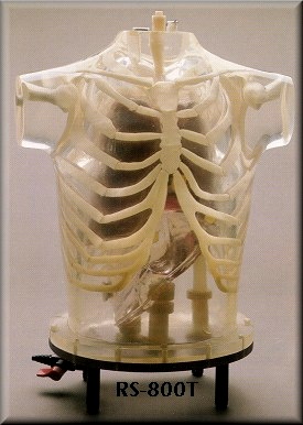
    The volume of each vacuum-formed breast is 972 ml. A tumor, filled with a radioactive solution can be inserted to evaluate the planar and tomographic imaging techniques used for mammoscintigraphy. A set of breast tumors 3, 6, 9, 12, and 15 mm diameters) is included. They are supported by thin, threaded nylon rods which pass through ports and are sealed by o-rings. They can be bent by hand to reach any part of a breast.

    The thorax is mounted on a base plate with an o-ring seal. Four rubber feet provide space under the phantom for drain fittings. The base plate is removed readily to provide access to the interior of the phantom.


    A knob at the top of the neck secures a rod which passes through to the heart/lung subassembly for disassembly.
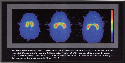
    STRIATAL PHANTOM FOR SPECT/PET
  • The Head
  • The Head Phantom is based upon a standard RDS Head with a calvarial cut to insert or remove the brain shell easily. The nasal vacity and maxillary sinuses are filled with foam with a mass density of 0.23 g/cc.

  • Brain Shell
  • The Brain Shell has five compartments which can be filled separately; left and right nucleus caudate, left and right putamen, and the remainder of the brain. This allows different nucleus caudate to putamen ratios as well as different striatal to background ratios to be obtained; this also permits differences between left and right striatal activity to be examined.

    The volume of the Brain Shell is about 1260 ml. The volumes of the nucleus caudate and putamen are 5.4 ml and 6.0 ml respectively.


    Quantification of striatal uptake is not straightforward because it depends on a number of factors:
    a) Type of radionuclide used (99mTc, I-123 or F-18)
    b) Imaging factors such as: collimator type, amount of scatter and attenuation
    c) Image processing parameters such as: scatter and attenuation-correction techniques, type of reconstruction filter, slice thickness, region-of-interest size and its location.

    In normal subjects, the putamen and head of the nucleus caudate are small structures with typical dimensions of 7-15 mm in the axial plane (that are comparable to the system resolution). Since partial volume effects are more important for objects with dimensions less than twice the system resolution, the selection of imaging and reconstruction parameters is critically important in calculating the striatal-to-occipital ratio used to measure the relative striatal uptake in the brain.


    Stock #
    Description
    Your Price
    RS-800T
    Heart/Thorax Phantom (includes RS-801 through RS-810)
    $17,730.00
    RS-801
    Thoracic Cavity with bottom place (price if sold separately)
    $10,240.00
    RS-802
    Non-perfusable lungs (pair)
    $1,550.00
    RS-803
    Perfusable Lungs withstyrofoam beads (pair) (price if sold separately)
    $1,700.00
    RS-804
    Heart with 3 hollow defects in myocardial wall-standard sizes (price if sold separately)
    $2,300.00
    RS-805
    Liver Shell-ONLY. (price if sold separately)
    $1,370.00
    RS-806
    Chest Overlay (price if sold separately)
    $1,370.00
    RS-807
    Removeable breast with set of tumors (price if sold separately)
    $1,640.00
    RS-809
    Set of 10 threaded nylon support rods (price if sold separately)
    $60.00
    RS-810
    Set of 5 external fillable markers (price if sold separately)
    $110.00
    RS-811
    Tumor ONLY, with rods, Specify size of tumor:
    3mm, 6mm, 9mm, 12mm, 15mm (price is per tumor)
    $310.00
    RS-900T
    Head with transparent brain shell containing striatum
    $5,787.60
    RS-901T
    Transparent brain shell with striatum
    $1,738.40
    RS-542
    Thyroid Phantom without Head; Consists of neck, hollow thyroid,
    cover, a portion of the cervical spine, sternum and clavicles
    $4,740.00
    RS-545
    Thyroid Phantom with head;
    same as RS-542 but with head and full cervical spine
    $5,510.00
    RS-543S-T
    Thyroid-hollow, shell
    $728.00
    RS-543A-T
    Thyroid-hollow, active
    $Call


    Cards We Accept!
    For order methods and policies, please click here.


    E-Mail Sales
    Telephone

    Phone Lines open...
    8am - 5pm Monday-Friday (Pacific/West Coast Time)
    Phone: 1-503-667-3000
    Toll-Free (US): 800-827-XRAY (800-827-9729)
    Download our order form
    and then...Fax Your Order
    24 Hours a day!
    1-503-666-8855

    P.O. Box 625 * Gresham, OR  97030 * U.S.A.

    This web site is our catalog, no printed catalog is available. - Please keep checking back, changes to this site are made daily.
    Every effort is made on our part to keep all posted prices up to date, however we can not guarantee the accuracy due to notification delays by the manufacturers.
    Purchase Orders accepted only upon approval of credit - Min. $500 opening order for new open CHARGE accounts.
    All other orders (Cash, Credit Card, etc) subject only to a $30 product minimum order!


    ©1997-2024 Pacific Northwest X-Ray Inc. - Sales & Marketing Division - All Rights Reserved